FLAME facility (Medical School)
Functional and Light-Activatable Multi-dimensional Electrophysiology facility
Who we are
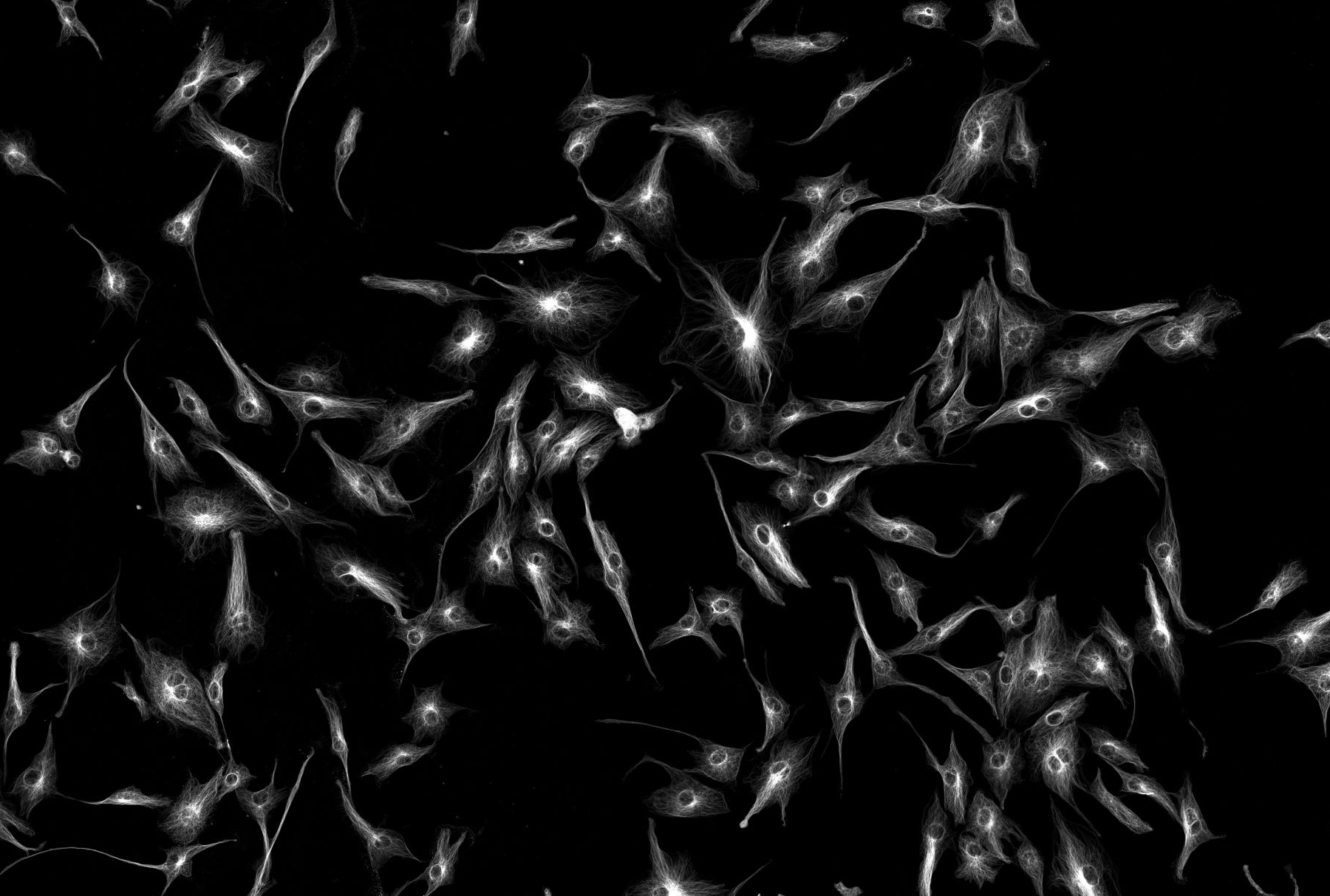
Micro-Electrode-Array (MEA) Lab
In 2020, a group of NUBI neuroscientists and stem cell biologists led by Prof. Evelyne Sernagor and the FMS Bioimaging Facility (Alex Laude) were awarded a BBSRC ALERT grant to develop a novel, cutting-edge experimental platform for in vitro investigation of neural network function at multiple scales and unprecedented spatial and temporal resolution.The platform combines electrical and optical recordings. The electrical recording component consists of a new-generation, large-scale, high-density multielectrode array (MEA) recording device system with pillar electrodes capable of recording from neurones deep within the tissue.This MEA is integrated with epifluorescence microscopy in order to combine electrical and optical measures of various physiological parameters. The platform can be used to record from a wide range of isolated preparations such as brain slices from various species (including human tissue), retinas and stem-cell derived organoids to investigate neural function in health and disease. It could potentially also be used to record from non-neuronal samples. Thanks to the penetrating electrodes, it is possible for the very first time to record deep in the tissue, thus allowing sampling of in vitro networks that is unparalleled in its extent and resolution. Combining this innovative approach with optical tools opens exciting new avenues of research, using a variety of optical biosensors for important physiological parameters and optical actuators to manipulate function. The platform is accessible to a wide range of internal and external users, including academics, early career researchers trained to use innovative technology, and industrial partners. It is available for national and international collaborations and offers unique opportunities to train a new generation of researchers. Moreover, it can be used as a testbed for the future development of new MEA chips and provides an invaluable tool for drug development and toxicology studies.
Neuromuscular Junction Research (NMJ) Lab
The Applied Neuromuscular Junction Facility is a research facility for confocal microscopy and electrophysiological applications. The primary aim of the facility is to investigate the structure and function of neuromuscular junction (NMJ) using mouse models -mimicking human diseases- as well as human motor point biopsies. The facility houses state-of-the-art imaging and electrophysiology equipment not only to allow researchers to investigate intracellular recordings of synaptic events from NMJs but also to capture high quality confocal images. In our facility you will have the opportunity to generate high resolution images of subcellular structures and molecular events with great sensitivity and high speed as well as detection of voltage recordings and ion channel currents in isolated nerve and muscle preparations. Moreover, the Facility is equipped with state-of-the-art patch-clamp devices permitting high-resolution recording of the ionic currents through the plasma membrane. We offer a range of services to meet your research needs, from the sample preparation, acquisition, analysis of multichannel fluorescence-based images and electrophysiology recordings to hands-on training in microscopy and electrophysiology.
Address
Cookson Building
Ground floor, room G.31
Dental School
The Medical School
Framlington Place
Newcastle Upon Tyne
NE2 4HH
UK
Contact us