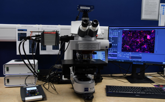
Zeiss AxioImager with Apotome (System 1)
Modalities: upright, widefield
Overview
The Zeiss AxioImager is a high quality wide field imaging system that offers three modes of operation:
- brightfield transmitted light (phase and DIC)
- wide field fluorescence
- apotome structured illumination (confocal like) fluorescence
The system utilises two separate camera systems for colour and monochrome imaging.
The Apotome (structured illumination) mode allows users to collect confocal-like images and ‘Z’ stacks, which can subsequently be projected as 3D images (stereo anaglyphs) and movies.
The illumination source is Colibri 7.
This system is capable of visualising Cy7 as it has the appropriate filter and wide spectrum lightsource.
The microscope has as an autofocus system and a precision motorised stage which enables users to automatically acquire tiled images from large specimens (or from an entire slide) and stitch them together post capture.
Analysis of data and images can be carried out using the Zen Blue software which includes quantification and colocalisation modules.
Specification
Here you can find the specifications for this microscope.
Objectives
Magnification |
NA |
Coverslip |
Other modes |
Immersion |
|---|---|---|---|---|
| 2.5 | 0.075 | Air | ||
| 10 | 0.3 | 0.17 | Air | |
| 20 | 0.8 | 0.17 | Air | |
| 40 | 0.75 | 0.17 | DIC | Air |
| 63 | 1.4 | 0.17 | DIC | Oil |
| 100 | 1.4 | 0.17 | Ph3 | Oil |
Filters (epi)
Cube |
Example fluorophores |
Excitation |
Dichroic |
Emission |
Part Number |
|---|---|---|---|---|---|
| DIC/Analyser | |||||
| Zeiss 112 | DAPI, AF488, AF555, AF647, Cy7 | 335-385 | 405+439+ 575 + 654 + 761 | PBP 425/30 + 514/31 + 592/25 + 681/45 + 785/38 | |
| Zeiss 38 HE | GFP, FITC, AF488 | 450-490 | 495 | 605/70 | |
| Zeiss 43 | Cy3, AF555 | 545/25 | 570 | 630/75 | |
| Zeiss 20 HE | TRITC, Rhodamine | 540-552 | 560 | 575-640 | |
| Zeiss 49 | DAPI | 365 | 395 | 445/50 | |
| Zeiss 50 | Cy5, AF660 | 640/30 | 660 | 690/50 | |
| Zeiss 91 | CFP, YFP, mCherry | 423/44 | 450+538+610 | TBP 467/24+555/25+687/145 |
*Discontinued for the newest SpOr-B-000 with a slightly different dichroich (FF506-Di03 instead of FF506-Di02)
Light source (epi)
Colibri 7 (385nm/430nm/475nm/555nm/630nm/735nm)
Detectors (cameras)
Flash4 camera
Zeiss MRc (Colour for brightfield HC)