Image Analysis
Our Image Analysis Unit
The Image Analysis Unit is made up of image analysis specialists with varying interests and backgrounds. We create custom, personalised, image analysis solutions for researchers and technical staff across the university. We have access to a wide range of image analysis software, and offer training and guidance in image analysis, including practical workshops and seminars on common tools such as ImageJ.
Our Team
George Merces – Image analysis specialist and team leader, develops open-source automated pipelines for image analysis. Has experience in wide range of image analysis source modalities, including light microscopy, electron microscopy, flow cytometry and adjacent technologies, medical imaging, and photogrammetry.
Jamie Grimshaw – Bacteriology specialist, providing image analysis support, expert bacterial experimental design guidance and advice, and assistance with microscopy image collection at CBCB.
Emma Foster – Bioimaging specialist with diverse imaging and analysis experience. Works with users from the BioImaging Unit to guide imaging protocols for optimum analysis conditions. Provides expert support in developing ImageJ pipelines for users from BioImaging and beyond.
Between us, we have experience in a range of scripting languages to help design automated image analysis solutions for research. We also provide training in more general image analysis modalities, including through the running of practical workshops and lectures.
Our Process
Our process always begins with a single question: What research questions are you hoping to have answered with this analysis? From there, we guide users on how to establish their experiments, put them in contact with the correct facility to perform their imaging, and begin designing a custom automated approach to answer their questions. We can get involved at any point in the research pipeline, whether that’s before you perform an experiment or once you’ve got your images and want to analyse them. We keep constant communication with our researchers so they remain in full control of their research, while we provide the technical and expert solutions to their problems. Validation of our automated approach is carried out with input from the researchers, so they can have confidence in the techniques we apply and have the knowledge to defend their work if needed. We assist with manuscript preparation so dissemination remains clear and easy for other researchers to replicate.
We also offer training in image analysis techniques for researchers who would prefer to learn how to do it themselves, with solo tutorials and group workshops giving practical skills to researchers at all experience levels.
Example of our work - 60 channel images
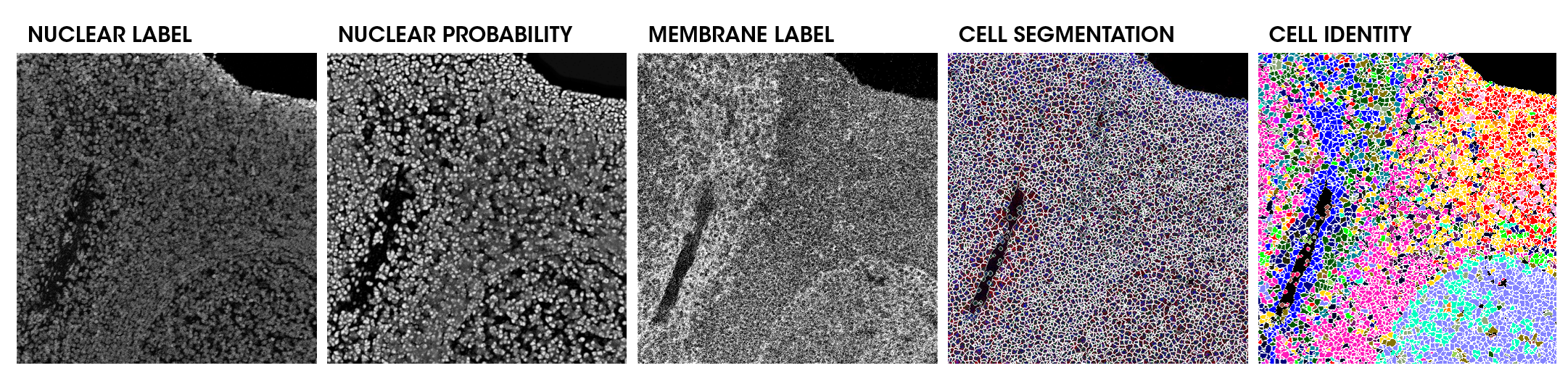
Here we have an example of taking the complex label information from channels (Nuclear Label and Membrane Label), to structures (Nuclear Probability) using complex machine learning tools, to single-cell segmentation using optimised pipelines (Cell Segmentation), all the way through to single-cell identification (Cell Identity). You can read more about this specific example here: https://www.biorxiv.org/content/10.1101/2023.02.21.526083v1