High-Content Imaging
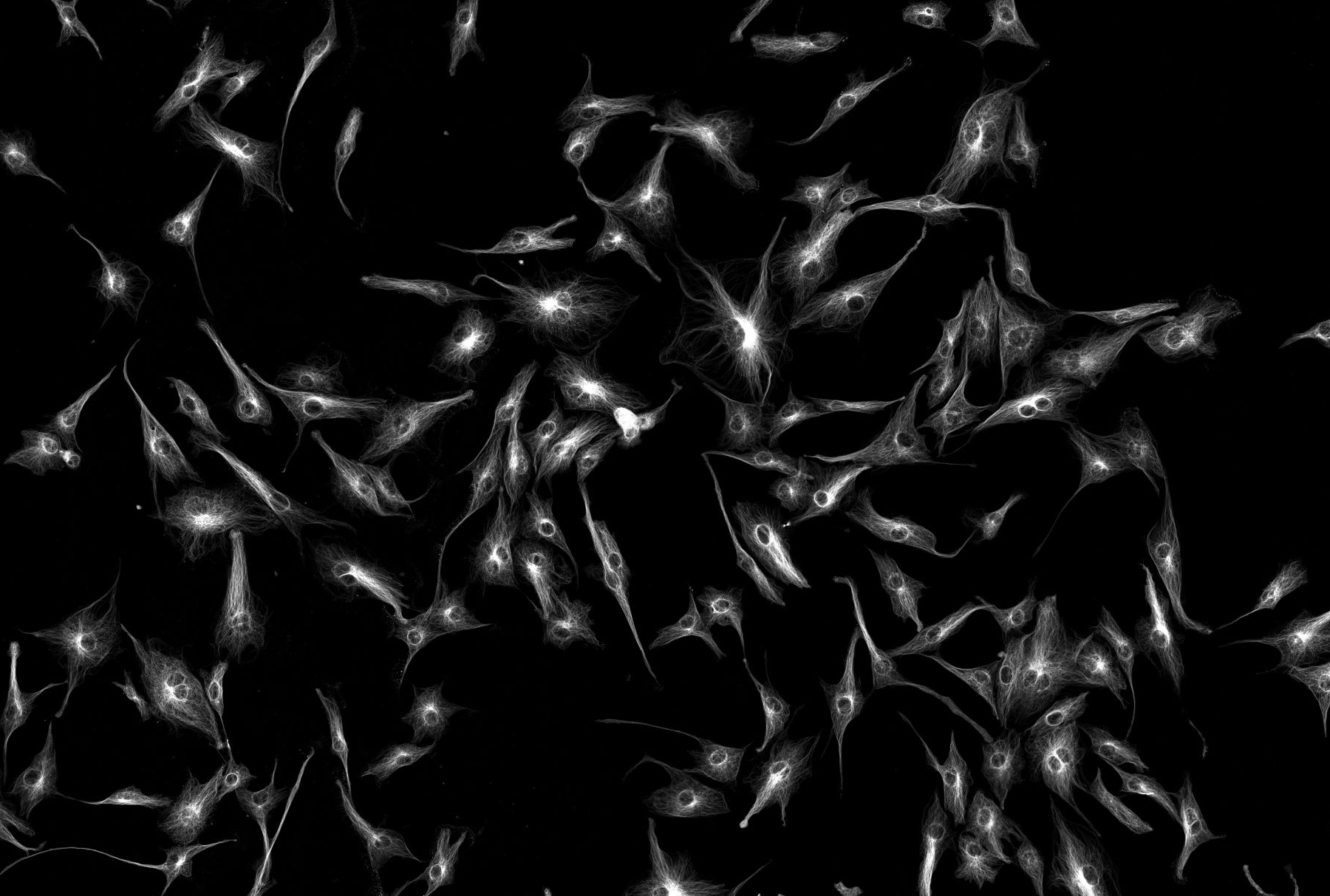
High content imaging takes the traditional microplate assay to the next level, allowing for the capture of high resolution widefield or confocal images of entire multiwell plates. This gives you the opportunity to screen multiple experimental conditions in an automated and repeatable manner. Where a plate reader will provide quantitative data from a population cell based assay, high content imaging will increase scope of data, to not only allow for quantitation of signal, but can also provide phenotypical and localisation data from the cells as well as population heterogeneity.
This approach is useful in screening large data sets from drug screens where the added information provided by the images may offer a greater insight into the effects of a specific compound on the morphology of the cell which a traditional microplate assay could not.
We offer a selection of different options for dealing with your automated large scale imaging needs, both in fixed cells as endpoint assays, or in live cells (with CO2 and temperature control) as single point or repetitive/time-lapse image profiles.
Our microscopes
Zeiss Cell Discoverer 7
Our flagship high content system running Zeiss’ Zen software suite is a fully automated widefield microscope system complete with 40 place room temperature and 40 place incubated plate storage system with integrated robotic loader and scheduling system. Using a combination of air and water immersion lenses and a multi wavelength LED illumination system the CD7 allows for automated multiplate and multi slide imaging captured on a highly sensitive Hamamatsu Fusion sCMOS camera. The system also allows for a conditional or guided acquisition experiment to be designed and automatically analysed, further increasing the automation possibilities for large scale plate or slide arrays.
Zeiss LSM800 AiryScan
Utilising the same tiles and positions module within Zeiss’ Zen platform, our inverted super-resolution confocal microscope offers environmentally controlled imaging of samples on a more traditional platform than the CD7. The LSM800 offers both traditional confocal and Airyscan super-resolution imaging on a fast stable easy to use platform.
Nikon TiE (1) – JOBS (invert)
A widefield more traditional style upright microscope, but fully automated allowing for widefield and colour imaging of microscope slide and multiwell plates in an environmentally controlled stagetop chamber. The system can be used for fixed or live cell imaging and runs Nikon Elements JOBS high content acquisition and analysis package
Nikon A1r+ resonant JOBS (invert)
Utilising a similar inverted TiE stand, but with a full microscope environmental chamber and outfitted with an A1R HD confocal scanhead/laserbed, this microscope can be used for live cell imaging in either Galvano or Resonant mode and utilising Nikon’s Denoise AI to provide high contrast images rapidly and with minimal bleaching. It also utilises Nikon Elements JOBS module to provide a high content acquisition and analysis platform, but adding the benefit of confocal volumetric imaging.
AxioScan7
Software Perkin Elmer Columbus with spotfire
Specialist high-content-focussed image analysis software.
IncuCyte Sx5
The Incucyte® Live-Cell Analysis System is designed to efficiently capture cellular changes where they happen - in the incubator. It is capable to capture high-resolution fluorescence and bright field images and record data in real time over hours, days or weeks.
