Our Facilities
We have a wide range of imaging facilities and expertise. The University facilities are all dedicated for full-time research investigation.
Clinical MRI facilities
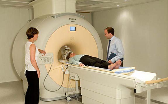
The Newcastle Magnetic Resonance Centre conducts research-driven imaging studies using our two Philips 3 Tesla clinical scanners.
The Magnetic Resonance (MR) physics team have strong collaborations with Philips medical systems. This allows our team to:
- programme novel MR acquisition and processing methods
- offer flexibility beyond traditional clinical imaging
From June 2012 the Newcastle MR Centre has engaged 115 studies, spanning basic and clinical research and commercial trial activity.
Methodology developed by the MR physics team is being used as primary or secondary clinical endpoints in three National Institute for Health Research (NIHR) Efficacy and Mechanism Evaluation Programme (EME) trials. Newcastle lead two of the trials and Manchester is leading the third.
A Stroke Association/British Heart Foundation funded clinical trial was led by Kings College, London.
The MR Physics Team is also a co-ordinating imaging for two multi-national trials. These trials are:
- the Jain Foundation study of Dysferlinopathy
- an EU FP7 study and clinical trial in brain iron storage diseases, “TIRCON”
