Services and Imaging Modalities
Computerised Tomography (CT)
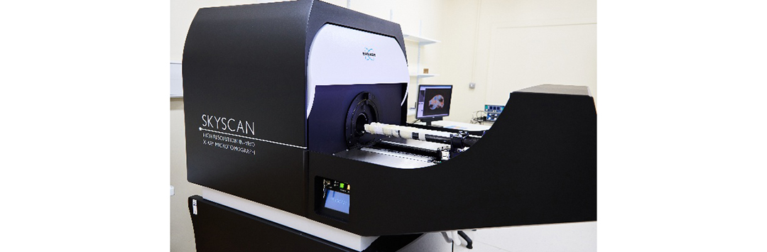
The PIVI facility is equipped with a high performance and resolution in vivo micro-CT system.
The SkyScan 1176 scanner provides an ideal anatomical imaging modality for non-invasive visualisation of lung, fat and bone, without the need for CT contrast agents. With its extra-large imaging bore and long axial field of view, the 1176 model offers excellent flexibility to image a wide range of subjects.
In material sciences, the 1176 system has provided engineering groups with a non-destructive method to investigate shape, porosity, density and other characteristics
Animal monitoring
The physiological monitoring sub-system includes video monitoring of an animal with real-time movement detection, ECG and breathing detection, and temperature stabilization.
In Vivo Imaging System (IVIS)
Funded by the Wellcome Trust, this 3D optical imaging system allows non-invasive and real-time longitudinal monitoring of disease progression, cell trafficking, and gene expression patterns. The system is capable of both luminescence and fluorescence modes of imaging.
Real-time in vivo imaging
The IVIS can image bioluminescence and fluorescence in up to 5 mice during a single scanning session. Images can be used to generate high quality 3D quantitative representations of bioluminescence and fluorescence (400–840 nm) in vivo and in real time.
The IVIS software allows for easy switching between fluorescence and bioluminescence modes and facilitates in vivo spectral unmixing of fluorescent probes.
The system also incorporates a topographic laser scanner, and can yield for single-view, diffuse tomographic reconstructions of internal sources.
IVIS techniques
- Bioluminescent
- Spectral un-mixing
- Fluorescence, including epi-illumination and trans-illumination
- 3D diffuse tomographic, including Fluorescent Imaging Tomography (FLIT) and Diffuse Luminescence Imaging Tomography (DLIT)
Potential applications of IVIS
- Functional genomics
- Expression profiles and regulation study
- Protein–protein interaction
- Apoptosis study
- Oncology
- Tumour growth and metastasis
- Tumour related gene study
- Infectious disease
- Infectious pathway
- Molecular study
- Stem cell research
- Tracking and functional analysis
- Phamaceuticals
- Drug discovery (high throughput)
- Pharmacokinetics (PK)
- Absorption, distribution, metabolism and excretion (ADME)
- Toxicology research
- Pharmaceutical toxicology
- Chemical toxicology
- Gene delivery and therapy
- Expression kinetics and localisation
Animal monitoring
IVIS is equipped with an Isoflurane anaesthesia system providing accurate flows of 0.5 and 1.0 L/min of gas (O2).
IVIS stage temperature can be monitored where the subjects are placed.

Bruker 4.7 Tesla Magnetic Resonance Imaging Centre for Translational Systems Neuroscience (CTSN)
The pre-clinical Centre for Translational Systems Neuroscience (CTSN) facility houses a Bruker 4.7 Tesla (T), 38 cm bore. This is ideally suited for vertical/upright scanning of behaving NHPs.
This scanner is a part of an international initiative working towards NHP MRI open data (PRIME-DE: Milham et al., Neuron, 2018; Milham, Petkov et al., Neuron 2020). We are a key site providing high-quality and high-resolution structural and functional MRI data in behaving NHPs as an indispensable translational model for understanding the human brain in health and disease. The scanner has also supported contracts, such as for scanning soft tissue at high resolution, and national and international collaborations.
MRI is an extremely versatile imaging modality that can be utilised to collect dynamic, functional and metabolic information in vivo. The 4.7T and 7T pre-clinical systems are also currently being developed for smaller NHP scanning, in collaboration with University of Cambridge.
MRI techniques
The 4.7T MRI scanner offers a broad range of imaging techniques including:
- Anatomic imaging (T1/T2/Proton-Density weighted imaging)
- Functional magnetic resonance imaging (fMRI)
- Diffusion-weighted imaging
- Resting-state fMRI
- Animal welfare monitoring
- Magnetic resonance spectroscopy (MRS)
- Blood oxygen measurements (BOLD EPI methods)
- High-resolution laminar-resolved fMRI
The 4.7T MRI is equipped with a range of specialised coils for pre-clinical imaging including:
- 4 and 8 channel phased-array imaging coils
- Array of transmit/receive coils
- GRAPPA reconstruction for protocols using parallel acceleration
- Presentation of auditory and visual stimuli
- Eye tracking system, lever press system, reward delivery system
- Flexible interface for experimental setups
Potential applications of MRI
- Neuroscience and neurodegenerative disorders
- Optogenetics effects visualisation (opto-fMRI)
- Diffusion tensor imaging (DTI)
- Deep brain stimulation visualisation (es-fMRI)
- Transcranial ultrasound neurostimulation (collaboration with University of Oxford)
- Musculoskeletal disorders
Contact
For further information on the Bruker 4.7T MRI system please contact Dr Balezeau
Shear Wave Elastography (SWE)
The PIVI facility benefits from a clinical grade Shear Wave Elastography (SWE) system.
SWE is a medical imaging technique used in ultrasound to assess the stiffness or elasticity of tissues. This non-invasive method provides valuable information about tissue characteristics and is particularly useful in the evaluation of liver fibrosis and other soft tissue abnormalities.
- Shear Wave Elastography (SWE)
- Tissue elasticity
- Doppler / Colour imaging
- Fluid motion – velocity & direction
- 2D B-mode anatomical imaging
- Structural measurement

Small Animal Radiation Research Platform (SARRP)
Small Animal Radiation Research Platform
The Small Animal Radiation Research Platform (SARRP) at Newcastle University represents a cutting-edge preclinical radiotherapy system designed to bridge the gap between laboratory research and clinical radiotherapy. This advanced system enables researchers to conduct image-guided radiotherapy studies with unparalleled precision, replicating clinical treatment workflows and predict radiotherapy response in patients.
The SARRP features a 225kV X-ray tube mounted on a 360-degree rotating gantry, paired with a motorised specimen stage to provide high-resolution Micro-CT imaging and precise radiotherapy delivery. Using the MuriPlan software, 3D CT data is utilised to outline and target the tumour while sparing healthy tissue. The SARRP's flexible field of view and adjustable collimators enable the simultaneous irradiation of multiple animals, as well as the irradiation of both 2D and 3D cell cultures.
Key Features of SARRP
Cone Beam CT (CBCT) Imaging
CBCT provides high-resolution anatomical images, ensuring accurate tumour and tissue localisation for precise targeting.
- High-throughput: Multiple animals can be treated at the same time.
- Quick acquisition: Images can be captured in under 1 minute.
- High resolution: 127 µm for detailed imaging.
Calibrated robotic specimen stage and X-ray gantry
The SARRP features a motorised specimen stage with full XYZ and theta controls, offering precise positioning. While the X-ray gantry provides 360 degrees of rotation around the system's isocentre. This unique calibration allows for the sequential targeting and treatment of multiple regions within the same animal with 0.2mm accuracy.
Customisable Beam Configurations
- Motorised Variable Collimator (MVC): The SARRP's MVC enables precise targeting of lesions or entire organs by dynamically adjusting the X-ray field size. Field sizes can range from a minimum of 2mm x 2mm in diameter to a maximum of 35 mm x 75 mm, adaptable from any angle.
- Fixed Collimators: The system includes six fixed collimators with sizes from 0.5 – 10mm (0.5mm, 1mm, 5mm, 10mm, 3x3mm, 3mm x 9mm, 10mm x 10mm). The fixed collimators provide versatility for various applications, allowing for quick and efficient beam size selection with minimal penumbra.
MuriPlan Software
Image reconstruction and treatment planning: MuriPlan enables the reconstruction of images from X-ray projections taken at 360-degrees. The resulting 3D image is used for precise treatment planning to delineate the target area while minimising exposure to surrounding healthy tissue.
Integrated Dosimetry Planning: Allows for real-time dose calculations, mimicking clinical radiotherapy planning software.
Integration with other imaging modalities: SARRP can be integrated with multiple imaging modalities, including micro-CT, optical, MRI, PET/SPECT, with image fusion tools to clearly identify the target and organs at risk.
Physiological monitoring and gating
- The SARRP is equipped with a respiratory gating module to reduce motion artefacts and enables precise X-ray beam delivery to small lung and abdominal lesions.
- The gating system allows continuous monitoring of animal breathing during both imaging and treatment.
- Heated beds are provided to maintain the animal's physiological temperature throughout the procedure.
- The system is fully integrated with an isoflurane anaesthesia setup, ensuring seamless anaesthesia delivery.
"Thanks to the team at the Newcastle Preclinical In Vivo Imaging Facility we can now add microCT to our repertoire of tools for post-mortem forensic investigation. The images that this technique produce can provide easily understood documentation of important findings that can be used to help us assist the police with their enquiries."
Tracy Sorkin & Nigel Cooper, NHS Forensic Sciences, Newcastle upon Tyne Hospitals NHS Foundation Trust
Training and Equipment Access
We are keen for individuals to access much of our equipment themselves, and can provide the training necessary to gain the required levels of competence and confidence for this to happen. The level of training required varies between each imaging modality and the type of work being performed.
A general introduction to the facility and its equipment is also provided to anyone requiring access, and covers all relevant safety and access procedures.
Equipment availability and booking can be accessed using our online booking calendar.