

Mega Microscope
One small step towards detecting Life on Mars
Published on: 13 October 2016
Biological evidence of life in basalt rocks - similar to those found on Mars - has been detected for the first time.
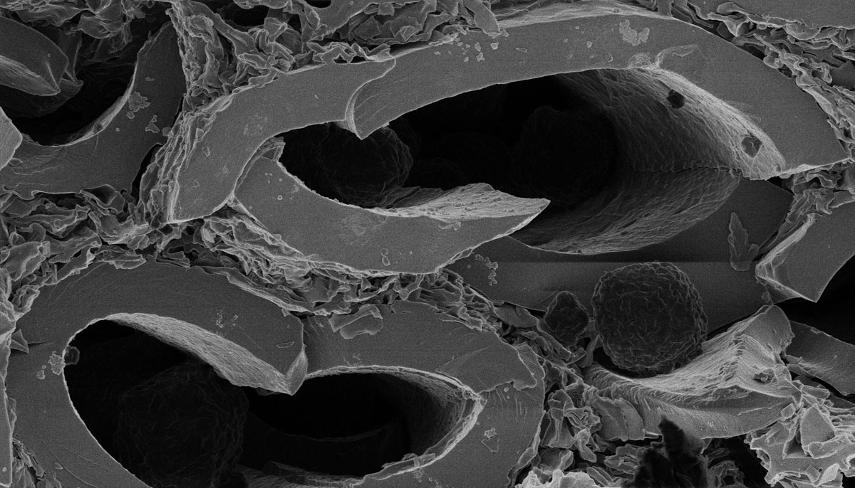
Using state-of-the-art technology, experts at Newcastle University, UK, have been able to identify a variety of organic compounds deposited in microscopic ‘tunnels’ in basalt rocks recovered from the Ontong Java plateau in the Pacific Ocean.
Believed to have been left behind as finger-like tubule structures made by microbes 200 million years ago, this is the first time that anyone has been able to provide such detailed analysis of the organic material which is likely of biological origin.
Analysed in Newcastle University’s new £7m Surface Engineering and Analysis Laboratory, the findings are an important step towards answering one of the most fundamental questions in science today: Are we alone?
Publishing their findings in the Journal of Vacuum Science and Technology, the next step is to analyse a piece of Martian rock, searching for the remnants of organic molecules left behind by ancient microbes.
Looking for extra-terrestrial life
Geoscientist Graham Purvis explains:
“Our findings provide the strongest evidence to date that these structures have a biological origin and because sub-oceanic basalt has very similar properties to that which we would expect to find on Mars it means we can use the same technique to look for signs of extra-terrestrial life.
“Over the years, there have been many claims made about evidence of early life on Mars, but all have been contentious.
“A key problem with the detection of extra-terrestrial life is that the origin of ancient rock structures can often be explained both biologically and abiotically - that is they could have been created by a living organism or by some chemical or physical action.
“The search for evidence of life on Mars is not an easy task because of the scarcity of pristine, uncontaminated Martian meteorite samples."
ToFSIMS and the Helium Ion Microscope
Newcastle University’s Surface Engineering and Analysis Laboratory, part-funded by the Engineering and Physical Sciences Research Council (EPSRC), will be officially opened today and is the only one of its kind in the UK with the capability to analyse surfaces to such a high degree of accuracy.
Combining the capabilities of the EPSRC National XPS Centre with the new equipment - ToFSIMS and a Helium Ion Microscope - the hope is the new facility will open the door on a number of new discoveries in health, engineering and science.
“The possibilities with this technology are both huge and tremendously exciting,” says head of the new laboratory Professor Peter Cumpson.
“What we have detected is the trace left behind by living systems - the biological fingerprint which even after 200 million years is still visible - albeit only with a very, very good microscope!”
ToFSIMS - or Time of Flight Secondary Ion Mass Spectrometry - detects very low concentrations of surface impurities. Measuring to a depth of just a few atomic layers, the sample surface is hit with a beam of primary ions (such as Oxygen). These remove material as secondary ions from the surface.
“The ToFSIMS measures the mass of these secondary ions and it is from this that we can deduce what the surface was made of,” explains Professor Cumpson.
“An uncontaminated surface will produce one pattern but if, for example, the sample is contaminated by an oil molecule then the mass of the secondary ions will be different."
The new £1.5m Helium Ion Microscope works in a similar way but due to the very short wavelength of the Helium ions it means they can be focussed to a much smaller spot, giving much clearer, higher-resolution images and providing a much greater level of detail. At Newcastle the He Ion Microscope is fitted with a SIMS analyser allowing 10nm resolution - five times better than available anywhere else in the UK.
Professor Cumpson adds:
“Techniques such as XPS have long been used in the electronics industry for fault-finding in semiconductor manufacture and processing.
“But the Helium Ion Microscope can be used for the first time for the analysis of soft materials - such as surface contaminations of needles, vials and medical implants, or for tracking drugs and where they end up in the body - so healthcare applications are growing strongly.”
Reference: Gas cluster ion beam for the characterisation of organic materials in submarine basalts as Mars analogues.
Naoko Sano, Graham Purvis, Anders Barlow, Geoffrey Abbott, Neil Gray and Peter Cumpson
Journal of Vacuum Science and Technology A, 2016 http://dx.doi.org/10.1116/1.4954940


Cutting Edge
Newcastle University's Time of Flight Secondary Ion Mass Spectrometry
Life on Mars?
He Ion Microscope image of fossilised Devonian/Silurian flora plant or fungus
State-of-the-art
Newcastle University's Time of Flight Secondary Ion Mass Spectrometry Surface Engineering and Analysis Laboratory


